Pre-Clinical Optical Imaging
Bioluminescence and Fluorescence Imaging
The IVIS Spectrum is a bioluminescence and fluorescence optical system for in vivo and ex vivo imaging. The magnitude of the optical signal allows for longitudinal, non-invasive assessment of targets of interest via expression of a bioluminescent/fluorescent reporter gene or administration of a fluorescent probe. A wide range of commercially available targeted fluorescent probes facilitate in vivo imaging of angiogenesis, perfusion, inflammation, apoptosis and bone turnover. In addition, fluorescent in vivo imaging agents have been designed that activate florescence in the presence of various disease-associated proteases, e.g. metalloproteinases and cathepsin. Furthermore, dyes for labelling peptides, proteins, nanoparticles and antibodies are available.
Accordingly, the applications afforded by this system are truly diverse & cover the key therapeutic areas such as cancer, cardiovascular disease, & inflammation as well as for novel drug evaluation studies in these therapeutic domains. Utility of this imaging system has already been proven, as shown via output publications using this technology.
Instrumentation IVIS Spectrum
Optical imaging is a relatively high-throughput technique compared with other in vivo imaging modalities, such as MRI or PET, given that up to 40 animals can be imaged per hour (5 simultaneously).
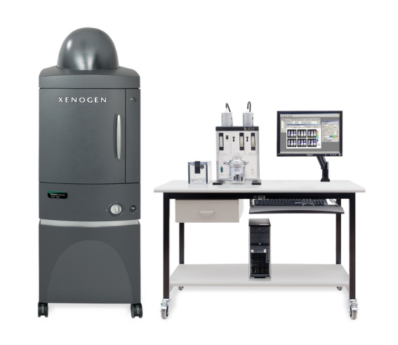
The IVIS Spectrum is a high through put, high sensitivity, low noise, optical imaging system with bioluminescence and fluorescence capabilities that includes:
- Cooled CCD camera (-90° C) mounted on a light-tight imaging chamber, cooling to -90° C facilitate very low level light imaging
- Integrated anaesthesia unit and a large field of allow 5 animals to be imaged simultaneously
- Broad range of excitation filters (10 excitation filters from 430nm to 745nm) and emission filters (18 emission filter from 500nm to 840nm)
- Broad band Fluorescence excitation Light Source
- Computer Control System
- High through put up to 40 animals per hour depending on protocol
- Living Image software for IVIS spectrum image viewing and data analysis is available on work stations in the BMF and on a dedicated imaging analysis computer in the Conway Institute area reading area 6.

